
When I first began reviewing granulation tissue wound pictures during hands-on wound care, I quickly learned that they tell a deeper story than most guides suggest. These images are not just snapshots—they are visual checkpoints that reveal whether the body is steadily progressing through the stages of repair or signaling a potential setback.
From my own observations, a wound that develops a moist, beefy-red tissue bed is usually on the right track, while pale, grayish, or friable tissue can indicate problems such as poor circulation or infection risk. Understanding these differences early allows patients and caregivers to intervene quickly and support faster, safer healing.
In this article, we’ll break down how to read granulation tissue wound pictures, highlight the key signs of progress, and share practical recovery tips drawn from real-world cases and research.
Top Takeaways
- Wound pictures show progress. They help confirm if healing is on track or delayed.
- Healthy tissue looks moist and red. Pale, uneven, or dark tissue signals a warning.
- Photography improves outcomes. Research shows patients heal better with visual documentation.
- Builds confidence and trust. Pictures provide visible proof of healing and motivate patients.
- Act quickly on changes. Always pair photos with professional medical advice.
What Wound Pictures Really Reveal
Granulation tissue wound pictures provide a clear visual of the body’s repair process.
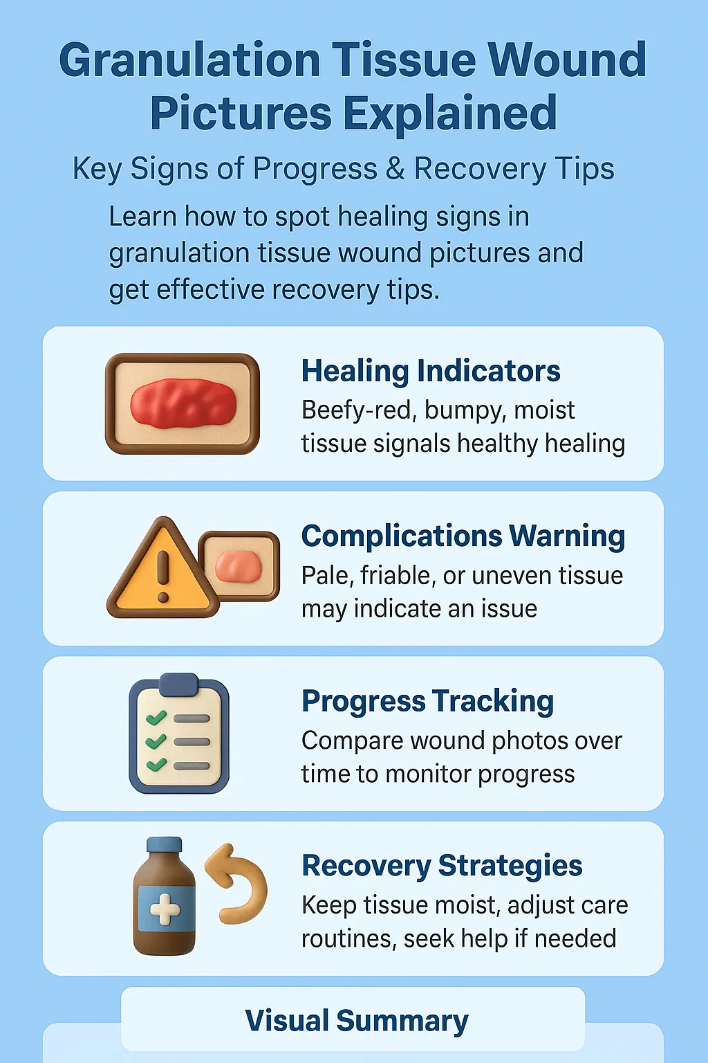
- Healthy signs: Red, moist, slightly bumpy tissue with good blood supply.
- Warning signs: Pale, gray, dry, or uneven tissue that could point to circulation problems, infection, or delayed healing.
By comparing images over time, both patients and caregivers gain insights into whether the wound is steadily rebuilding or needs medical intervention, while also monitoring the presence and type of exudate as an important indicator of healing progress.
Expert Insight
"In my experience, granulation tissue wound pictures are more than clinical references—they’re progress markers. A healthy red tissue bed signals rebuilding, while pale or uneven tissue often reveals setbacks. Learning to interpret these details can be the difference between smooth recovery and prolonged complications."
Case Studies & Real-World Examples
Post-Surgical Healing
Patient: 54-year-old after abdominal surgery
Initial signs: Pale, uneven tissue → poor circulation
Interventions: High-protein diet, improved dressings, increased mobility
Outcome: By week 4, photos showed healthy red tissue
Takeaway: Pictures reassured both patient and care team that healing was on track
Chronic Diabetic Foot Ulcer
Patient: Long-standing ulcer in a diabetic patient
Challenge: Slow healing, unhealthy friable tissue
Interventions: Debridement and infection management
Outcome: Transition to healthy red granulation within weeks
Takeaway: Regular photos guided timely decisions and boosted patient motivation
Research Perspective
Evidence shows documenting wounds with photos improves outcomes and helps patients understand their healing journey
First-hand experience: Photos are teaching tools that empower patients and enhance clinical care
Patient: 54-year-old after abdominal surgery
Initial signs: Pale, uneven tissue → poor circulation
Interventions: High-protein diet, improved dressings, increased mobility
Outcome: By week 4, photos showed healthy red tissue
Takeaway: Pictures reassured both patient and care team that healing was on track
Patient: Long-standing ulcer in a diabetic patient
Challenge: Slow healing, unhealthy friable tissue
Interventions: Debridement and infection management
Outcome: Transition to healthy red granulation within weeks
Takeaway: Regular photos guided timely decisions and boosted patient motivation
Evidence shows documenting wounds with photos improves outcomes and helps patients understand their healing journey
First-hand experience: Photos are teaching tools that empower patients and enhance clinical care
Supporting Statistics
Chronic wounds are widespread
8.2 million Medicare patients are affected annually, costing $28–$96 billion
Source: National Institutes of Health – Chronic Wounds Study (nih.gov)
Insight: Early changes spotted in wound pictures can prevent costly complications.
Pressure ulcers are common
Over 2.5 million people develop them annually in the U.S.
Source: Agency for Healthcare Research and Quality – Pressure Ulcer Facts (ahrq.gov)
Insight: Photos help caregivers catch tissue breakdown before it worsens.
Diabetic foot ulcers are high-risk
12–25% of people with diabetes will develop a foot ulcer in their lifetime
Source: National Library of Medicine – Diabetic Foot Ulcers (nih.gov)
Insight: Regular wound pictures reassure patients by showing visible progress.
Long-term challenges are severe
Recurrence rate: up to 65% within 5 years, with high amputation and mortality risks
Source: American Diabetes Association – Foot Complications (diabetes.org)
Insight: Teaching patients how to interpret wound photos gives them control and improves adherence.
Clinical guidance supports photography
Recognized as a valuable addition to written records
Source: National Library of Medicine – Wound Photography Guidelines (nih.gov)
Insight: Photos often reveal subtle tissue changes missed in written notes.
Final Thought & Opinion
Granulation tissue wound pictures are far more than medical records—they are windows into recovery. They make invisible progress visible, provide patients with reassurance, and help clinicians catch problems early.
In my experience, pictures often succeed where words fall short: they show healing, motivate patients, and reveal subtle setbacks before they become serious. My opinion is simple—granulation tissue wound pictures are essential tools for recovery, trust, and better outcomes.
Next Steps
Compare your wound: Red and moist = progress; pale or gray = warning
Track healing: Take consistent photos (same lighting, angle, time)
Consult professionals: Share photos with your provider for expert guidance
Use reliable resources: NIH, CDC – Diabetes Complications, ADA
Act fast if needed: Seek help immediately if pictures reveal infection or stalled healing


